A tissue culture experimental model is best for to study the direct effect of diabetogenic chemicals not possible products of its metabolisation on pancreatic B-cells. Authors are adopted to tissue culture model of isolated pancreatic islets histological and histochemical methods analysis of histostructure of islets and of insulin content in B-cells. The high quality results of analysis of histostructure of islets were obtained using Aldehydefuchsine method and most precise results of estimation of insulin content in B-cells by using of fluorescent Pseudoisocyanine and Immunofluorescent methods.
In vivo model of experimental diabetes caused by injection or by oral administration of diabetogenic chemicals is not objective regarding obtained results: very often it is difficult to estimate does morphological changes in pancreas are determined by direct alterative effect of diabetogenic substances or this effect is caused by other metabolites formed in organism as result of metabolisation or transformation of diabetogenic injected chemicals in liver, blood or in gastro-intestinal system. Is not possible to know what concentration of injected diabetogenic substance is delivered to pancreatic islets by blood.
Using of experimental model of isolated by Collagenase pancreatic islets it is possible to investigate direct effect of various concentrations of diabetogenic chemicals on pancreatic islets structure and on state of pancreatic B-cells. It is important advantage of model in vivo which possess to obtain objective data about direct action of investigated substances on pancreatic B-cells. Meanwhile not all histological and histochemical methods widely used for staining of p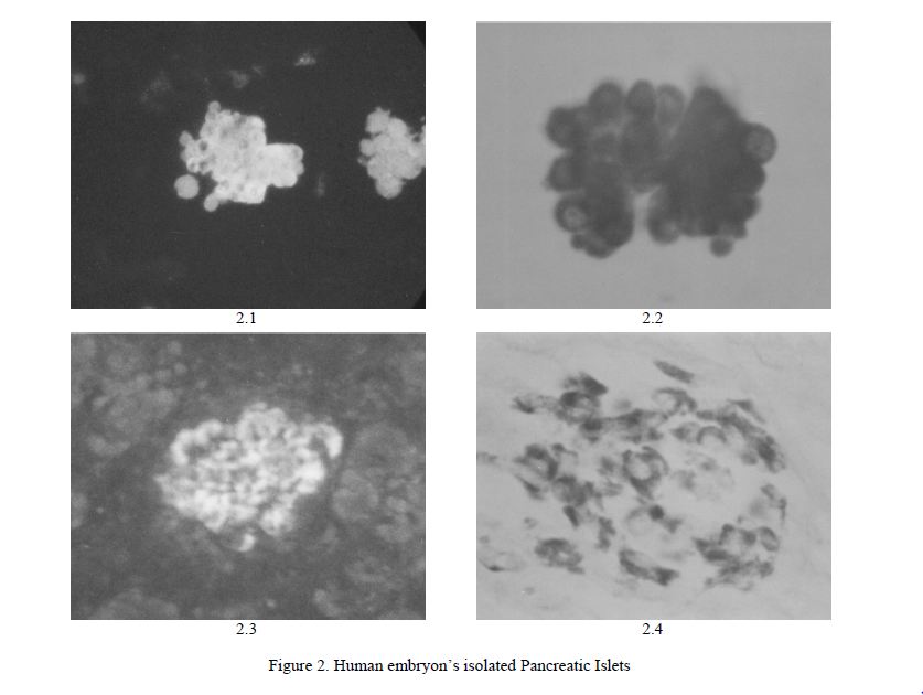
Aim of work: to adopt methods of fixation and staining procedures for using of isolated pancreatic is lets.
Materials and methods
Animals. Pancreas of 24 rats LEWIS 4–5 days old and 8–10 weeks old human embryons were used.
Isolation procedures: dissected pancreas tissue were treated 3 times 3 min each by 2 % solution of Collagenase (Boehringer Mannheim, Germany; FLUKA, Switzerland); rinse 3 times in cold Hanks solution and centrifugation; cultivation 12h at +37º Celsius in medium RPMI 1640 (SERVA, Germany) with bovine serum + 5.5 mM of Glucose, pH 7.32–7.38. Fixation in Bouin 15 min – 1 h and filling in paraffin. Sections 4 mcm were used for staining. After deparaffinisation sections were stained by methods: aldehydefucshin (basic fucshin from Avocado Chemical company, USA and MERCK, Germany) [1], immunofluorescent method (antiserum for insulin from Institute of Diabetes «Gerhardt Katsch», Germany), Victoria 4R method (FERAK, Germany, MERCK, Germany) [2], Die thylpseudoisocyanine method (SERVA, Germany) [3] and Haematoxylin and Eosin method. All methods were adopted for isolated pancreatic islets tissue [4–6].
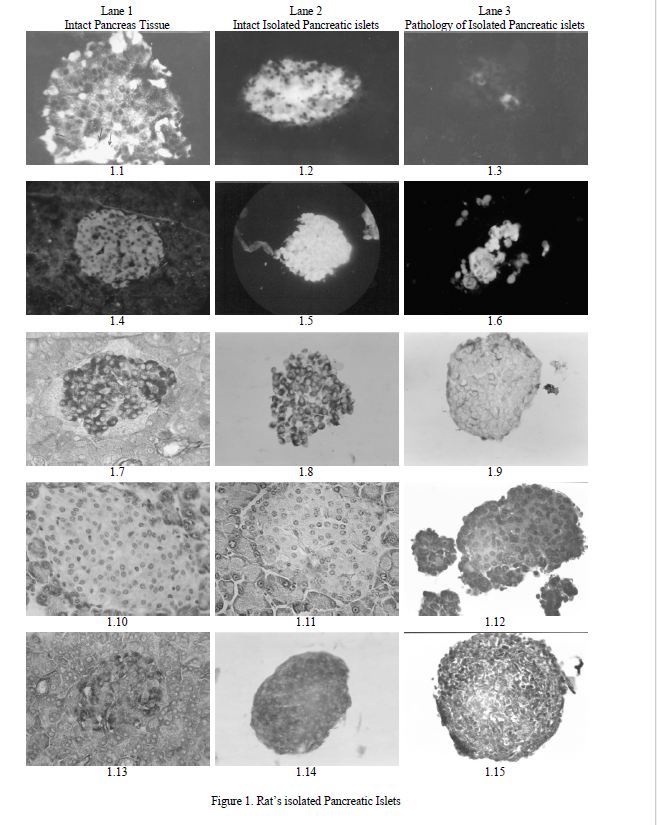
Figure 1. Rat’s isolated Pancreatic Islets
- Frozed section of fresh islet without staining; ×280;
- Positive reaction for Insulin. Immunohistochemical method; ×160;
- Negative reaction for Insulin. Immunohistochemical method; ×160;
- Positive fluorescent reaction for Insulin in B-cells on Pancreas tissue; Diethylpseudoisocyanine; ×140;
- Positive fluorescent reaction for Insulin in B-cells of isolated Islet; Diethylpseudoisocyanine; ×140;
- Destruction of isolated Islet and negative fluorescent reaction for Insulin in B-cells; Diethylpseudoisocyanine; ×140;
- Positive reaction for Insulin in B-cells on Pancreas tissue; Aldehydefucshine; ×280;
- Positive reaction for Insulin in B-cells of isolated Islets; Aldehydefucshine; ×280;
- Destruction of isolated Islet and negative reaction for Insulin in B-cells; Aldehydefucshine; ×280;
- Islet in Rabbit’s Pancreas; Haematoxylin and Eosin; ×280;
- Islet in Rat’s Pancreas; Haematoxylin and Eosin; ×280;
- Isolated Islet of Rat’s Pancreas; Haematoxylin and Eosin; ×280;
- Islet on Pancreas tissue; Victoria 4R; ×280;
- Positive reaction for Insulin in isolated Islet; Victoria 4R; ×280;
- Negative reaction for Insulin in isolated Islet; Victoria 4R; ×280;
Figure 2. Human embryon’s isolated Pancreatic Islets
- Pancreatic Islet in study of formation; positive reaction for Insulin; Diethylpseudoisocyanine; ×140;
- Pancreatic Islet in study of formation; positive reaction for Insulin; Victoria 4R; ×370;
- Pancreatic Islet in study of formation; positive reaction for Insulin; Immunofluorescent method; ×140;
- Pancreatic Islet in study of formation; positive reaction for Insulin; Aldehydefuchsine; ×140. Microphotographs of histological sections:
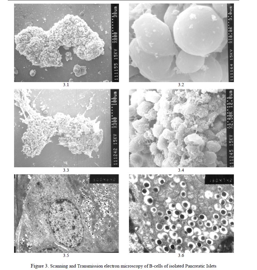
Figure 3. Scanning and Transmission electron microscopy of B-cells of isolated Pancreatic Islets
- Isolated intact islet. Scanning electron microscopy; ×310;
- Isolated intact islet. Smooth surface of individual A-cells on the surface of islet. Scanning electron microscopy;
- Isolated islet. Destruction of cells. Scanning electron microscopy; ×320;
- Destruction of isolated Damaged surface of individual A-cells on the surface of Islet. Scanning electron microscopy; ×540;
- Isolated intact Multiple B-granules contained Insulin; cell matrix without changes. Transmission electron microscopy; ×3150;
- Multiple B-granules in B-cells of isolated islet; cell matrix without changes. Transmission electron microscopy;
Scanning electron microscopy method. Drying of islets past incubation by CO2 spraying by gold and were investigated in scanning electron microscope Hitachi S-570 at using voltage 15 kV. Transmission electron microscopy method. Past cultivation in medium RPMI 1640 islets were fixed in 2 % Glutaraldehyde solution 30 min. Filling of Islets in Durcupan. Sections were investigated on JEM-7A electron microscope.
Results
Immunofluorescent staining method [IF]. We have obtained same results of staining by IF as using of sections of pancreas tissue (Fig. 1.1–1.3). IF is high specific method for revealing of Insulin in B-cells. Decreasing of Insulin content in B-cells of islets past action direct action of Streptosotozin was evidently demonstrated by this method (Fig. 1.3).
Diethylpseudoisocyanine chloride fluorescent method [PS], a high specific for revealing A-chair of molecule of Insulin, showed same result comparatively using of sections of Pancreas tissue (Fig. 1.4–1.6). Time for staining of sections in 0,4 % solution of Diethylpseudoisocyanine was reduced from 20 min. till 15 min. as was reduced time for washing of sections past staining procedures. This method showed marked decreasing of Insulin content in damaged B-cells (Fig. 1.5, 1.6) in compared with intacts.
Aldehydefuchsine method showed analogical results (Fig. 1.7–1.9). A significant differences are revealed of state of histostructure as of Insulin content in damaged isolated islets comparatively with intacts (Fig. 1.8, 1.9). Aldehydefucshine method [AF] contrary to IF and PS is not belong to high specific because colours other hormones too. But for pancreatic B-cells not contained other hormones AF is specific for Insulin.
Haematoxylin and Eosin method using we obtained same result in compared with using sections of Pancreas tissue (Fig. 1.10–1.12.).
Staining of human embryon’s pancreatic B-cells
Human embryon’s 8–10 weeks old is not formed completely yet and is as small or more large groups of B-cells like small islets (Fig. 2.1–2.4.) contained a large amount of Insulin revealed by all histochemical methods used by us. Diethylpseudoisocyanine technic and Immunofluorescent method as fluorescent more sensitive methods demonstrated very positive reaction for Insulin in B-cells (Fig. 2.1, 2.3). Staining by Victoria 4R method showed intensive diffuse staining of cytoplasm of B-cells (Fig. 2.2). Aldehydefuchsine technic showed not intensive staining of B-cells (Fig. 2.4).
Scanning and Transmission electron microscopy analysis of isolates pancreatic islets
Pancreatic islets of 4–5 days old LEWIS Rats have oval or irregular shape (Fig. 3.1.). Surface layer of islet formed by A-cells which have spherical or oval shape (Fig. 3.2). A-cells have smooth surface (Fig. 3.2.). Islets treated by Diphenylthiocarbazone, a diabetogenic chelat active chemical, looked like the islets with clear signs of damage and destruction (Fig. 3.3.) and with evident damage of surface layer formed by A-cells (Fig. 3.4).
Transmission electron microscopy analysis showed results similar to the observed in the study of endocrine pancreas tissue. Cell matrix of B-cells of isolated islets as ultrastructures of cells without changes and contained multiple B-granules contained Insulin (Fig. 3.5, 3.6.)
Discussion
Analysis of results showed that using of histological and histochemical methods for staining of sections of isolated pancreatic islets have similar or equal to similar results obtained in pancreas tissue past staining by same methods. Fluorescent histochemical methods as Immunofluorescent reaction for Insulin as method using of Diethylpseudoisocyanine are more sensitive and identify the very low concentrations of investigated substances as 10–7–10–8, that has been confirmed by our results. Meanwhile both these methods have a common fault: histological sections past completing of staining procedures are not permanent and must be investigated within short time. Both methods are belong to high specific for staining of Insulin or of A-chair of molecule of Insulin. These methods are more precise for measuring intensity of insulin staining in B-cells because no other structure of islets are stained.
More suitable for practical using is Aldehydefucshine technic. Histological sections of pancreas tissue as of isolated islets stained by this method are permanent and can be stored for a long time. Aldehydfuchsine method is not belong to high specific for Insulin staining. It is known that some pituitary hormones can also be stained by Aldehydefuchsine method. Meanwhile for pancreatic islet’s B-cells this method you can be measured as specific for insulin because the other hormones in B-cells are not synthesized. Method Victoria 4R is high specific for Insulin and as Aldehydefuchsine technic gives an opportunity to obtain permanent histological sections. Quantitative estimation of insulin content in stained sections is based on measuring of absorbed by B-cells of light. However, both of these methods are belong to histological methods too and result staining not only of Insulin, but also other structures of B-cells which absorbed light as Insulin. Therefore, results of estimation of Insulin content in the B-cells by measuring of absorbance is not so precise as using fluorescent histochemical methods for Insulin staining.
We used significantly reduced time for fixation of Islets in Bouin from 24 h for pieces of pancreas tissue up to 15–30 min. for isolated Islets. Time for staining of sections of isolated islets by Diethylpseudoisocyanine was reduced to 15 min. comparatively with 20 min. for sections of pancreas tissue.
Acknowlegement
Authors are thankful for financial supporting of this investigation to Prof. John Turtle, a Honorary VicePresident of International Diabetes Federation (IDF) (Sydney, Australia), to Victor Riedl (Wien, Austria) and for free reagents to Dr Hans Langisch, Director of Eastern Europe Division of the Boehringer Mannheim GmbH (Mannheim, Germany), to Dr Hans Niedderer, President of SERVA Finebiochemica GmbH (Heidelberg, Germany), Dr. Elizabeth Horn (Heidelberg, Germany), Dr. Johannes Watzek (Mannheim, Germany), Dr Peter Felch and Dr Robert Schilly (Wien, Austria), to Prof. Frank Wohlrab (Leipzig, Germany), Prof. Bernard Tuch (Sydney, Australia), Dr Harald Ritzel (Frankfurt, Germany), Dr Konrad Niethammer (Darmstadt, Germany), Dr Ch.Studer (Zurich, Switzerland) and to Dr Roland Thunberg (Uppsala, Sweden). Authors are thankful for help to Doz. G.T.Tusupbekova and Doz. A.P.Andreewa (Karaganda).
References
- Kvistberg , Lester G., Lazarow A. Staining of Insulin with Aldehydefuchsine // J. Histochem. & Cytochem. — 1966. — Vol. 14. — P. 104–111.
- Kikui , Segushi H., Mizuguti H. A differential staining method for Aand B-cells in the pancreas islets of Langerhans // Acta Histochem. & Cytochem. — 1977. — Vol. 10, No. 1. — P. 10–13.
- Coalson R.E. Pseudoisocyanin Staining of Insulin and Specifity of Emperical Islet Cell Stain // Stain. Technologies. — No. 2. — P. 121–129.
- Meyramov G., Meyramova R.G. The high Specifical Fluorescent Method Revealing of Zinc-ions in Pancreatic B-cells // Diabetes, the Journal of American Diabetes Association. — 1991. — Vol. 40, No. 5. — P. 65.
- Meyramov G.G., Kikimbaeva A.A., Meyramova A.G. Victoria 4R Method Staining of Insulin in B-cells of Isolated Pancreatic Islets // Acta Diabetologica, the European Diabetes — Springer International, 2003. — Vol. 40, No. 4. — P. 208–209.
- Meyramov G.G., Kikimbaeva A.A., Meyramova A.G. Fluorescent Histochemical method Staining of Insulin in B-cells of Isolated Pancreatic islets by Diethylpseudoisocyanine Chloride // Acta Diabetologica, the European Diabetes Journal. — Springer International, 2005. — Vol. 42, No. 1. — P.