Novel synthetic cannabinoids pose serious health risks to the society. One of the most significant concerns is constant appearance of new synthetic cannabinoids on grey markets. These substances cause numerous toxic effects in human body, ranging from neurotoxic effects to cardiovascular toxicity and acute kidney injury as well as addiction and withdrawal syndromes.
Thus, the aim of this study was to investigate cytotoxic effects of novel synthetic cannabinoids, THJ-018, on human neuronal cell line. The work reports the cytotoxicity of novel synthetic cannabinoid, THJ-018, on neuroblastoma, SH-SY5Y cell line, which has not yet been published in the scientific literature.
To study cytotoxicity of novel synthetic cannabinoids, SH-SY5Y neuroblastoma cell line was chosen to model cytotoxic effects of THJ-018 on neuronal cells in concentration of 5х10л4 cells/ml in vitro. THJ-018, dissolved in DMSO, was tested in concentrations from 1.0 to 100.0 цМ during 24 hours of exposure. To investigate cell viability, 3-(4,5-dimethylthiazol-2-yl)-2,5-diphenyltetrazolium bromide (MTT) assay was used. To count the number and percentage of viable cells, trypan blue (TB) cell counting assay, and Neubauer chamber were employed.
THJ-018 did not affect the viability of SH-SY5Y cells after 24h exposure in cell culture in vitro in the concentration range from 1.0 to 75.0 цМ. The results of MTT and TB assays showed that THJ-018 had no statistically significant cytotoxic effect on SH-SY5Y cells in terms of formazan production by mitochondria and percentage of viable cells in cell culture in vitro at these concentrations. However, at THJ-018 concentration of 100.0 цМ, production of formazan decreased from 100% to 71.5%± 11.8% if compared to negative control. The results of TB assay after 24h exposure to THJ-018 were in a good accordance with the previous results of the MTT assay. THJ-018 showed no cytotoxic effects on cellular viability in terms of the percentage of viable cells at concentrations from 1.0 to 75.0 цМ. But, at the concentrations from 75.0 to 100.0 цМ, percentage of viable SH-SY5Y cells in cell culture in vitro dropped from 100% to 67.3±15.6% and to 49.5±16.6% correspondingly, if compared to negative control.
The results of the study confirmed previously published reports that parent compounds of synthetic cannabinoids are less toxic than their secondary metabolites and smoke products in the concentration range from 1.0 to 100.0 цМ. However, at higher concentrations, cytotoxic effects become evident in terms of general cellular viability of mitochondria and percentage of viable cells.
The results of the work contribute to further understanding of mechanisms of cytotoxicity of synthetic cannabinoids in vitro.
Introduction.
13.3 % of young population (15-34 years of age) and 6.6 % of older population (15-64 years of age) used cannabis in 2017, whereas the usage of novel psychoactive substances (NPS), including synthetic cannabinoids (SCs), among this groups of population were only 3.0% [1].The importance of a more extensive research on toxicity of SCs is explained by a more toxic effect of their secondary metabolites than parent compounds, which is not observed in the case of the consumption of natural cannabinoids. Moreover, with two new SCs on the grey market every month, better understanding of neurotoxicity, protein and genetic toxicity of novel SCs, and study of their metabolism are needed for application in clinical studies and forensic investigations [2].
Pharmacological action of SCs includes psychotropic and immunotropic effects depending on the interaction with cannabinoid receptor (CBR) 1 or 2. SCs exert their psychotropic effects through CBR1 which is primarily located in central nervous system (CNS). Mechanisms of pharmacological action of SCs in CNS involve overstimulation of CB1 receptor, disruption of GABA/glutamate and endogenous cannabinoid system in the brain, re-uptake of CB1 receptor, activation of mitogenactivated protein kinase shown in forebrain and hippocampal neurons [3]. Immunotropic effects of SCs are caused by their binding to CBR2 which is mainly located in the immune system and the pharmacological action includes the relief of neuropathic pain and treatment of inflammatory diseases shown in human peripheral leukocytes, follicular and marginal B cells [4].
Toxicity of SCs was shown on body, organ and cellular levels. For instance, cardiovascular effects of toxicity of SCs include: tachycardia, hypertension, hyperglycaemia, and hypokalaemia. Other widely reported type of toxicological complication of the consumption of street drug products containing SCs is acute kidney injury (AKI). Clinical manifestations of such toxicity include electrolyte changes in urine and serum, evidence of hemorrhages in tissue from kidney biopsy demonstrated with different analytical methods including light microscopy, immunofluorescence, and electron microscopy [5].
Neurological effects are euphoria, drowsiness, paranoia, delusions, hallucinations, anxiety, panic attacks, agitation, nausea, vomiting, seizures and dizziness. Neurotoxicity can be manifested in cognitive defects and short-term memory loss and suicides [6]. SCs have much greater affinity to CBR1 than natural cannabinoids, as tetrahydrocannabinol (THC) in cannabis plant material, which makes them more potent psychoactive substances for central nervous system. Then, smoke products of novel SCs, such as smoke products of THJ--018, found in “Spice” street herbal drug product, were shown to exhibit cannabimimetic activity in mice. Finally, products of smoking of JWH-018 were also reported to retain psychoactive effects due to induction of CYP1A2 in the lungs and subsequent production of psychoactive metabolites in drug users [7, 8]. Moreover, psychotropic effects of SCs can be furthermore potentiated by their high lipophilicity and ability to cross blood-brain barrier and efficient distribution in the brain [9].
Unlike the mechanism of pharmacological action of SCs, the mechanism of body, organ, and cellular toxicity of SCs is not completely understood. Cellular and molecular toxicity of SCs includes damage to nuclear and mitochondrial DNA and cell membrane; alteration of genetic expression and protein synthesis, activation of caspase 3, apoptosis and necrosis [10]. For instance, the effects of a SC, JWH-018, in CBR1-expressing CHO cells in vitro, include inhibition of cyclic adenosine monophosphate production (AMP), whereas, in HEK293 cells expressing CBR1, JWH-018 activates mitogen-activated protein kinase and causes internalization of CBR1. Finally, in cultured hippocampal neurons, JWH-018 can inhibit excitatory postsynaptic neurotransmission in a dose-dependent manner [11].
Mechanism of toxicity of metabolites of SC, JWH-018, was described as independent of cannabinoid 1 and 2 receptors and targeting cell membrane integrity. According to the studied literature source, JWH-018 does not show a significant change on cell viability in HEK293T and SH-SY5Y cell lines. However, JWH018 N (3 -hydroxylated) metabolite caused the decrease in cell viability and necrosis in HEK293T and SH-SY5Y cell lines [12]. The mechanism of cytotoxicity of JWH-018 and other SCs was studied observing nuclear division index, formation of binucleated cells and nuclear abnormalities such as micronuclei, nucleoplasm bridges and buds, which could be indicative of toxic effect of SCs on the structure of the nucleus, chromatic packaging of DNA and nuclear division mechanisms, increase in the amount of DNA in the “tail” in single cell gel electrophoresis, which indicated single and double strand breaks in the DNA, shown in human buccal cells (TR-146), human lung-derived fibroblasts (A459) and human peripheral leukocytes [13].
Thus, the aim of this study was to investigate cytotoxic effects of novel synthetic cannabinoid, THJ-018, on human neuronal cell line, SH-SY5Y, by observing cell division and viability using trypan blue assay and mitochondrial redox metabolism by MTT assay.
Materials and Methods.
Cell Culture.
Cell culture in vitro was performed using human neuroblastoma cell line, SH-SY5Y, which was a gift from Prof. Dr. Tiago Outeiro, PhD (Institute of Molecular Medicine, Lisbon, Portugal). The cells were cultured under standard conditions (37°C, humidified atmosphere, 5 % CO2) in T25 cell culture flasks using DMEM which was supplemented with 10.0 % (v/v) bovine fetal serum and 1.0 % Pen/Strep-Glutamine. The SH-SY5Y cell line, which had been stored in liquid nitrogen, was recovered and used for the experiments after re-culturing from 3rd to the 11th passage.
The cells were passaged each time, when the confluence of the cell culture in T25 cell culture flask reached 70-80%. During cell passage, first, DMEM media was removed from T25 flask. Then, the cells were washed with 3.0 ml of Dulbecco's phosphatebuffered saline (DPBS). Next, DPBS was removed and the cells were detached with 1.0 ml of Triple Express, washed with 3.0 ml of DMEM, transferred into a 15-ml Falcon tube and centrifuged in the 15-ml Falcon tube at 1000 rotations per minute for 5 min. Finally, the solution was decanted and the cell pellet was resuspended in 3.0 ml of DMEM. This solution was used for passaging, cell counting and seeding of 96-well plates for the conduction of MTT and TB assays.
TB Assay.
Cell counting using TB was conducted according to Strober, W., 2001 with the following modifications [14]. 10.0 |iL out of cell suspension of total volume of 3.0 ml were transferred into a well of a 96-well plate and thoroughly mixed with 40.0 |iL of already prepared TB solution using a 20 and 200-^L pipettes achieving 1:5 dilution of cell suspension. After that, 10.0 |iL of this mixed solution were dispensed to fill-in a Neubauer chamber covered with a cover slip. This procedure was repeated twice to fill-in both chambers. Cells were counted at x40magnification in three big squares (central, upper left and lower right) and total concentration of cells in the volume of the initial solution of 3.0 ml of DMEM, in which the cell pellet was resuspended, was determined according to the following formula: concentration of cells (cells/ml) = the average of the number of cells in three big quadrants* 5.0 (dilution factor) * 104.
Counted cells were examined under the light microscope (x40) and determined either as viable cells in case they had clear cytoplasm, or damaged and dead cells in case they had blue cytoplasm. Finally, the percentage of viable cells was calculated according to the formula: % of viable cells = number of viable cells / total number of cells (viable and dead cells) / 100%.
MTT Assay.
MTT assay was performed according to the protocol given by Meerloo et al. (2011) with several modifications [15]. MTT assay measures cellular viability in terms of mitochondrial production of formazan crystals from MTT reagent. First, SHSY5Y cells were seeded into a 96-well plate at the concentration of 4.5 x 10⁴ cells/ml in DMEM in the volume of 200.0 |iL per each well and cultivated for 24h. Then DMEM was removed and the solutions containing the parent compound, THJ-018, were added at THJ-018 concentrations of 1.0, 5.0, 10.0, 25.0, 50.0, and 100.0 цМ.
THJ-018 solutions were prepared as follows. THJ-018 was dissolved in DMSO and then diluted in DMEM so that its concentrations were 1.0, 5.0, 10.0, 25.0, 50.0, and 100.0 цМ and the concentration of DMSO was always 0.2 % in the final volume of 200.0 |iL, which was added into each well of a 96-well plate. Each concentration was tested in 6 wells of the same column achieving 6 replicates, except for the columns designated for negative and positive controls. The cells were incubated with THJ-018 solutions at indicated concentration range for next 24 h. Next, DMEM was removed and the cells were washed with fresh DMEM in the volume of 100.0 |iL per each well. After that, MTT solution was added in the volume of 200.0 ^L per each well. The MTT solution (MTT powder from the stock dissolved in DMEM at the concentration of 0.5 mg/ml) was prepared fresh shortly before each MTT test. Aluminum foil was used to protect MTT solution from light. 0.2 % DMSO was used as negative control, whereas 1.0 % TritonX-100 was used as positive control.
The 96-well plate was unloaded by inverting it over and pressing it against paper towel in sterile conditions inside the biosafety 2 laminate hood. After that the cells were incubated with MTT solution for 2.5 h. Glycine buffer was not added at the end of the incubation. After the 2,5h incubation with MTT DMEM solution, the 96-well plate was unloaded as described above but in nonsterile conditions. Formazan crystals were resuspended in 100.0 % DMSO in the volume of 200.0 ^L per each well. The solution was thoroughly mixed with 200.0 |iL pipette, avoiding the production of bubbles which could interfere with absorbance reading and protected from light with aluminum foil. Finally, after formazan crystals were completely dissolved in 200.0 |iL of DMSO in each well, the plate was read at 595 nm using a Bio Rad 680 Microplate Reader.The results of MTT assay were given as percentage of the production of formazan in SH-SY5Ycells in cell culture in vitro in a well of a 96-well plate if compared to the negative control values, fixed as 100.0 %, from the same 96-well plate. Cellular production of formazan was directly related to the absorbance of the solution of formazan crystals dissolved in DMSO.
Results:
MTT Assay.
individual MTT assays, using SH-SY5Y cells from passages 3, 5, 7 and 9, were conducted to measure cytotoxic effects of the parent compound, SC, THJ-018, in the concentration range of 1.0, 5.0, 10.0, 25.0, 75.0, and 100.0 |iM, in these cells, measured in terms of the amount of mitochondrial production of formazan crystals from MTT reagent which is directly related to cellular viability.
As it can be inferred from the figure 1, parent compound of SC, THJ-018, did not affect the viability (formazan production) of SHSY5Y cells after 24h exposure in cell culture in vitro in the concentration range from 1.0 to 75.0 |iM. Conducted t-tests also showed no statistically significant differences between negative control and other datasets, except for the dataset on formazan production at THJ-018 concentration of 100.0 |iM, which was statistically significantly different from the negative control.
MTT Assay
Viability of SHSY5Y cell after 24Һ exposure
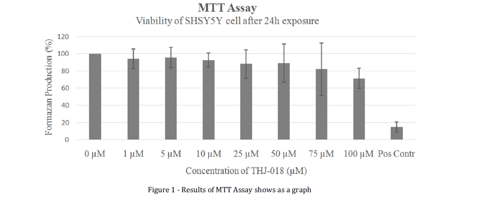
Figure 1 Results of MTT Assay shows as a graph
Results of MTT Assay, which measures the production of formazan crystals from MTT by cells through oxidationreduction metabolism in mitochondria, shows a graph of concentration of THJ-018 in цМ versus percentage of formazan production in % of negative control, which equals 0.0 цМ. For each data set, experiment was repeated 5 times (N=5), chisquare tests were used to compare data, P value of less than 0.05 was considered to indicate statistical significance, and standard deviation was calculated from the mean value using Excels Program. All P values are two-tailed.
At THJ-018 concentration of 100.0 цМ, production of formazan decreased from 100.0 % to 71.5 % ±11.8 % if compared to negative control.
MTT Assay
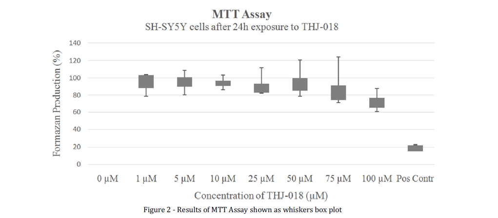
Figure 2 Results of MTT Assay shown as whiskers box plot
Results of MTT Assay, which measures the production of formazan crystals from MTT by cells through oxidationreduction metabolism in mitochondria, shown as a whiskers box plot of concentration of THJ-018 in цМ versus percentage of formazan production in % of negative control, which equals 0.0 цМ. For each data set, experiment was repeated 5 times (N=5), chi-square tests were used to compare data, P value of less than 05 was considered to indicate statistical significance, and standard deviation was calculated from the median value using Excels Program. All P values are two-tailed.
Constructed whiskers box plot of the results of MTT assay given in the figure 2 showed increase in the spread of values for production of formazan mostly above the median from lower to higher concentrations of THJ-018, which could be attributed to researcher error during the performance of MTT assays as well as to the conditions of cell culture during the exposition to higher concentrations of THJ-018.
TB Assay.
4 individual TB cell counting assays, using SH-SY5Y cells from 3, 5, 7 and 9th passages, were performed to measure the number of cells per ml after 24h exposure to THJ-018 in the concentration range of 1.0, 5.0, 10.0, 25.0, 50.0 and 100.0 цМ in cell culture in vitro. 0.2 % DMSO was used as negative control and 1.0 % TritonX-100 was used as positive control. Percentage of the number of cells was determined as percentage of the number of cells if compared to the negative control and measured with TB cell counting assay. The results of TB assay shown in the figure 3 indicate statistically significant increase in the number of cells up to about 130.0 % at 10.0 цМ and decrease to about 50.0 % and 30.0 % at 75.0 and 100.0 цМ of THJ-018 respectively.
Trypan Blue Assay
Figure 3 Results of trypan blue assay shown as a graph
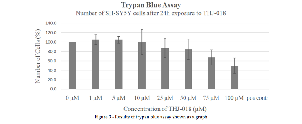
Results of trypan blue assay, which measures the percentage of viable cells versus dead cells by inclusion of Trypan blue dye, shows a graph of concentration of THJ-018 in цМ versus percentage of formazan production in % of negative control, which equals 0.0 цМ. For each data set, experiment was repeated 5 times (N=5), chi-square tests were used to compare data, P value of less than 0.05 was considered to indicate statistical significance, and standard deviation was calculated from the mean value using Excels Program. All P values are two-tailed.
Finally, the same results on the number of SH-SY5Y cells upon exposure to THJ-018, shown as a whiskers box plot.
Trypan Blue Assay
Number of SH-SY5Y cells after 24 h exposure to THJ-018
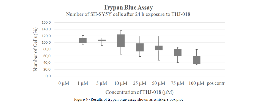
Figure 4 Results of trypan blue assay shown as whiskers box plot
Results of trypan blue assay, which measures the percentage of viable cells versus dead cells by inclusion of Trypan blue dye, shown as whiskers box plot of concentration of THJ-018 in цМ versus percentage of formazan production in % of negative control, which equals 0.0 цМ. For each data set, experiment was repeated 5 times (N=5), chi-square tests were used to compare data, P value of less than 0.05 was considered to indicate statistical significance, and standard deviation was calculated from the mean value using Excels Program. All P values are twotailed.
The figure 4 revealed a wide scatter of individual data points above and below the median in datasets on the number of SHSY5Y cells corresponding to THJ-018 concentrations of 10.0, 25.0 and 50.0 цМ, which could be explained by researcher error as well as differentiative conditions of cells in vitro under these concentrations of THJ-018 due to different passages and the phenomenon of hormesis.
Discussion.
The results of 4 conducted MTT assays after 24h exposure of SHSY5Y cells in 96-well plate in cell culture in vitro to the parent compound, THJ-018, did not show cytotoxic effect of SC, parent compound, THJ-018, at concentrations from 1.0 to
75.0 цМ in neuroblastoma cell line, SH-SY5Y. THJ-018 decreased viability of SH-SY5Y cells only at concentration of 100.0 цМ. As a result, viability of SH-SY5Y cells, measured as formazan production by MTT assay, fell from 100.0 % to 71.5±11.8 % if compared to negative control values.
As it could be observed from the figures 3 and 4 on the results of TB assay after 48h exposure of SH-SY5Y cells to THJ-018 parent compound, the graph of the percentage of number of viable cells formed a plateau on 130.0 % at THJ-018 concentrations from 1.0 to 10.0 цМ. This effect of the parent compound, THJ-018, could be explained by the effect of hormesis of SC, THJ-018, at these concentrations of neuroblastoma cell line, SH-SY5Y, resulting in stimulatory or beneficial effects instead of toxic or inhibitory effects of SC, parent compound, THJ-018. For example, the effect of hormesis of pesticides and drugs on biological systems, including animals, plants and cell cultures in vitro, was given by Calabrese and Baldwin, 2002 [16].
According to the theory of hormesis, upon exposure to SC, parent compound, THJ-018, at concentrations of 30.0 % of toxic level of 100.0 цМ, that is at THJ-018 concentrations from 10.0 to 50.0 цМ, SH-SY5Y cells in cell culture in vitro adapt to these concentrations of THJ-018 due to physiological overcompensation mechanisms leading to stimulatory effects of THJ-018 on cellular viability of SH-SY5Y cells and resulting in higher number of viable cells if compared to the negative control. Moreover, stimulatory and neuroprotective effects of SCs on neuronal cells, resulting in their higher proliferation rate in vivo and vitro, as well as better histological outcome of the brain tissue in terms of delay or reversal of death of neurons were shown by Calabrese, 2008 and Nagayama et al., 1999, in the context of research on rehabilitation pharmacotherapy after global or focal cerebral ischemia in in-vivo models [17, 18].
The nontoxic effects of the parent compound of SC, THJ-018, at concentrations from 10.0 to 50.0 цМ, could also be explained by the general mechanism of toxicity proposed in Couceiro et al., 2016, where cytotoxicity of SCs was described on the basis of SC, JWH-018, stipulating nontoxic effects of the parent compound versus toxicity of its secondary metabolites. Moreover, cytotoxic mechanism of action of secondary metabolites was shown as independent of CBR1, targeting, instead,the stability and integrity of cellular membrane.
Thus, the results of TB assay after 24h exposure to parent compound, THJ-018, were in a good accordance with the previous results of the MTT assay. The parent compound, THJ018, showed no cytotoxic effects on cellular viability in terms of the percentage of number of viable cells at concentrations from 1.0 to 75.0цМ. However, at the concentrations of parent compound, THJ-018, of 75.0 and 100.0 цМ, percentage of number of SH-SY5Y cells in cell culture in vitro dropped from 100.0% to 67.3±15.6% and 49.5±16.6% correspondingly, if compared to negative control.
Conclusion.
Thus, this present study, using MTT and TB assays, revealed that parent compound, THJ-018, did not show any statistically significant cytotoxic effects on cellular viability SH-SY5Ycell line in terms of formazan production and percentage of viable cells at concentrations up to 100.0 цМ.
The obtained results were in a good accordance with the previous publications on cytotoxicity of parent compounds of synthetic cannabinoids, which state that, because these parent compounds have not yet undergone metabolism, they are less reactive and cause less damage to cellular membranes and the nucleus. On the other hand, the results of the work also support the theory of hormesis, which explains absence of toxic effect of pesticides and drugs on biological systems, including animals, plants and cell cultures in vitro by adaptation to chronic stress. However, the results of TB cell counting assay of viable cells under chronic cell culture stress, such as sub-toxic concentration of synthetic cannabinoid, THJ-018, in cell culture medium during 24h, should be met with caution as the actual number of nonviable cells could be underestimated. For instance, the selectivity of cell toxicity assays, based on counting of the number of viable cells, was reported to be lower than the selectivity of molecular based assays, such as Annexin V antibody detection of apoptotic proteins, due to interfering factors of cell colony growth with significantly varying number of apoptotic and necrotic cells in each viable or toxicity assayed colony, which, in fact, was observed, in this present study, during TB cell counting of viable and nonviable cells after 24h exposure to parent compound, THJ-018.
Recommendations for future work.
This work is one of the first research attempts of the Laboratory of Molecular Pathology of Prof. Dr. Quintas at Egas Moniz Institute to assess neurotoxic effects of synthetic cannabinoid, parent compound, THJ-018, using SH-SY5Y cells as a model in vitro. The results of this study could be used in future neurotoxic assessment of parent compounds of synthetic cannabinoids in ever-evolving grey market of street drugs. Moreover, the findings, reported in this article, stress further research on viability and proliferation rate of neuronal cells under chronic cytotoxic stress of novel synthetic cannabinoids, such as parent compound, THJ-018, in terms of changes in genetic expression, cellular morphology, and mechanisms of apoptosis or necrosis, in order to further our understanding of neurotoxic effects of novel synthetic cannabinoids.
REFERENCES
- European drug report: trends and developments //Luxembourg: European Union: The European Monitoring Centre for Drugs and Drug Addiction (EMCDDA). 2017.
- Zawilska J. B., Andrzejczak D. Next generation of novel psychoactive substances on the horizon-A complex problem to face //Drug and Alcohol Dependence. 2015. Т. 157. С. 1-17.
- Koller V. J. et al. Toxicological profiles of selected synthetic cannabinoids showing high binding affinities to the cannabinoid receptor subtype CB1 //Archives of toxicology. 2013. Т. 87. №. 7. С. 1287-1297.
- Ashton J. C. et al. Cannabinoid CB1 and CB2 receptor ligand specificity and the development of CB2-selective agonists //Current medicinal chemistry. 2008. Т. 15. №. 14. С. 1428-1443.
- Buser G. L. et al. Acute kidney injury associated with smoking synthetic cannabinoid //Clinical toxicology. 2014. Т. 52. №7. С. 664673.
- van Amsterdam J., Brunt T., van den Brink W. The adverse health effects of synthetic cannabinoids with emphasis on psychosis-like effects //Journal of psychopharmacology. 2015. Т. 29. №. 3. С. 254-263.
- Wiebelhaus J. M. et al. Inhalation exposure to smoke from synthetic “marijuana” produces potent cannabimimetic effects in mice //Drug and alcohol dependence. 2012. Т. 126. №. 3. С. 316-323.
- Su M. K. et al. Metabolism of classical cannabinoids and the synthetic cannabinoid JWH-018 //Clinical Pharmacology & Therapeutics. 2015. Т. 97. №. 6. С. 562-564.
- Castaneto M. S. et al. Synthetic cannabinoids pharmacokinetics and detection methods in biological matrices //Drug metabolism reviews. 2015. Т. 47. №. 2. С. 124-174.
- Fantegrossi W. E. et al. Distinct pharmacology and metabolism of K2 synthetic cannabinoids compared to A9-THC: mechanism underlying greater toxicity? //Life sciences. 2014. Т. 97. №. 1. С. 45-54.
- Ferk F. et al. Genotoxic properties of XLR-11, a widely consumed synthetic cannabinoid, and of the benzoyl indole RCS-4 //Archives of toxicology. 2016. Т. 90. №. 12. С. 3111-3123.
- Couceiro J. et al. Toxicological impact of JWH-018 and its phase I metabolite N-(3-hydroxypentyl) on human cell lines //Forensic science international. 2016. Т. 264. С. 100-105.
- Bileck A. et al. Impact of a synthetic cannabinoid (CP-47,497-C8) on protein expression in human cells: evidence for induction of inflammation and DNA damage //Archives of toxicology. 2016. Т. 90. №. 6. С. 1369-1382.
- Strober, W. (2001). Trypan Blue exclusion test of cell viability. C u r r e n t P r o to c o l s in Im m u n o lo g y . DOI: 10.1002/0471142735.ima03bs21.
- van Meerloo J., Kaspers G. J. L., Cloos J. Cell sensitivity assays: the MTT assay //Cancer cell culture. Humana Press, 2011. С. 237-245.
- Calabrese E. J., Baldwin L. A. Defining hormesis //Human & experimental toxicology. 2002. Т. 21. №. 2. С. 91-97.
- Calabrese E. J. Dose-response features of neuroprotective agents: an integrative summary //Critical reviews in toxicology. 2008. Т. 38. №. 4. С. 253-348.
- Nagayama T. et al. Cannabinoids and neuroprotection in global and focal cerebral ischemia and in neuronal cultures //Journal of Neuroscience. 1999. Т. 19. №. 8. С. 2987-2995.