This review has considered some of the developments that have taken place with and which are shaping digital pathology. Of the different technologies surveyed, digital imaging is the one that will be adopted at the fastest rate. Today software programs enable pathologists to create and read the digital «slides.» Unlike traditional histological samples, microscopic users are able to create digital images almost any tissue sample traditionally viewed under a microscope. The advantages offered by cloud computing also allow researchers to create accessible databases and to share images rapidly. In outlining current trends, the chapter has described many of the advantages that arise from digitalization. The process remains a developing arena, however and there remain barriers to adoption. Barriers to adoption include limiting technology, image quality, problems with scanning all materials (e.g., cytology slides), cost, digital slide storage, inability to handle high- throughput routine work, regulatory barriers, ergonomics, and pathologists' reluctance. In time, many of these will be overcome as technology and education move on. Some possibilities of digital technology are facilitating personalized medicine where therapies are tailored to the individual and also empowering people to manage their own health through access to electronic health records. How quickly these come to pass remains to be seen; the key message is, however, that digital technology is transforming healthcare and the future delivery of pathology services in new and remarkable ways.
Introduction
The field of pathology continues to advance, along with other biomedicine fields, shaped by the advent of new technologies and practices. If investment can be taken as a signal of the pace of change, a report issued in 2017 put the worldwide market for digital pathology solutions at $5.7 billion [1]. Many of these developments are central to the digital transformation of the pathology laboratory. This chapter provides an overview if recent developments.
For pathologists the computer could soon become the diagnostic workstation, customized by the scientist to provide the resources to improve diagnostic outcomes, advise on treatments and monitor patient responses. While medical science is a specialist area it is not immune from the pressures faced by other workplaces. This takes the form of an increasing number of tests, and corresponding demand for faster, more- accurate disease diagnosis. These requirements, coming from both hospital managers and patients, can be better met through embracing digital technology. At the forefront of this is pathology. Pathology services play an important role in the study of disease, principally:
- Pathologists diagnose our illnesses;
- Pathologists screen us for congenital diseases, cancer and other conditions; and,
- Pathologists monitor the progress of disease and manage our therapies.
The vast majority of pathology laboratories still undertake manual procedures to reach a medical conclusion: these are tissue-based examinations performed using the traditional process of analyzing glass slidesamples using a microscope. However, it is possible for much of this process to be automated [2]. While new developments are taking shape, there are some barriers that need to be overcome. Some of the obstacles to adoption are also considered in this chapter.
In assessing the transformation of pathology, this chapter considers emerging processes like artificial intelligence; techniques for cell manipulation; digital plate counting, among others. To exemplify how digital pathology can be put into practice, the chapter closes with a brief case study review of the U.K. National Health Service.
The digital process
Before looking at some specific examples of digitalization, what are the main advantages for the pathology field? Digital technology has the potential to reduce subjectivity, improve diagnostic confidence and lead to more diagnoses being reproducible (an important step in reducing the risk of misdiagnosis). In terms of how these benefits have been taken and turned into practical and usable transformative technologies, examples of the digital transformation of pathology underway include:
- The scanning of histology slides;
- The use of analytical software (instead of personnel) to correlate patient data and images;
- The electronic delivery of patient information;
- Sharing of digital information between hospitals, including having another pathologist provide a second opinion rapidly (so-called «telepathology»);
- Having secure data transmission to avoid security breached;
- Setting up files of images from past cases so that diagnoses can be cross-compared;
- Establishing databases to assist with making medical decisions more quickly.
A few of these developments are considered below. Technological advances are also allowing for integration of the management of laboratory test results across multi-site labs using different lab information systems, such as hematology, immunology, microbiology, cytology and biochemistry. Consolidation through system integration can result in increased efficiency in processing data and supporting the function of multidisciplinary teams.
Artificial intelligence
Since the middle part of the second decade of the twenty-first century, several artificial intelligence systems have been launched within the laboratory marketplace. These are systems aimed at aiding the pathologist in making faster and more accurate decisions in relation to disease diagnosis.
One area where artificial intelligence and machine learning are set to make an impact is with the detection of cancer. This is a big step-change, considering that for over 150 years the classic approach to pathology has involved a sample of tissue being examined under an optical microscope, with the pathologist drawing upon his or her experiences to determine whether the tissue sample is cancerous.
New developments are changing this. For example, digital whole slide imaging enables the pathologist to capture an entire tissue sample on a slide. The image can then be digitally captured and analyzed with the help of specialized digital pathology software. Such software, such as that developed by Dr. David West Jr., of the imaging company called Proscia, allows laboratories to send images between each other, so that different opinions can be obtained. The digitalization also enables the software to perform part of the analysis. Here image analysis algorithms can engage in immunohistochemistry quantification. This process, whereby the algorithm can help to interpret the material, improves standardization and the consistency of decision making. It also speeds up analysis.
The use of artificial intelligence and machine learning means that the deep neural networks of the diagnostic machine can be trained to recognize broad or specific patterns on a whole slide image. The software can also interpret features in the tissue and make prediction (such as metastasis and recurrence). Where cancer is present, this can also be classified by the software (in terms of staging, grading, and differential diagnosis). This works on the basis of the digital image being interpreted by the machine in terms of numbers and the pattern of numerical variations is examined.
As technology has advanced many barriers to adoption have been removed, such as concerns with limiting technology, image quality, problems with scanning all materials digital slide storage, and ergonomics. Through multiple screening the artificial intelligence becomes better and the ability to spot predictive biomarkers becomes better, based on precise measurements of histological patterns.
Digital imaging
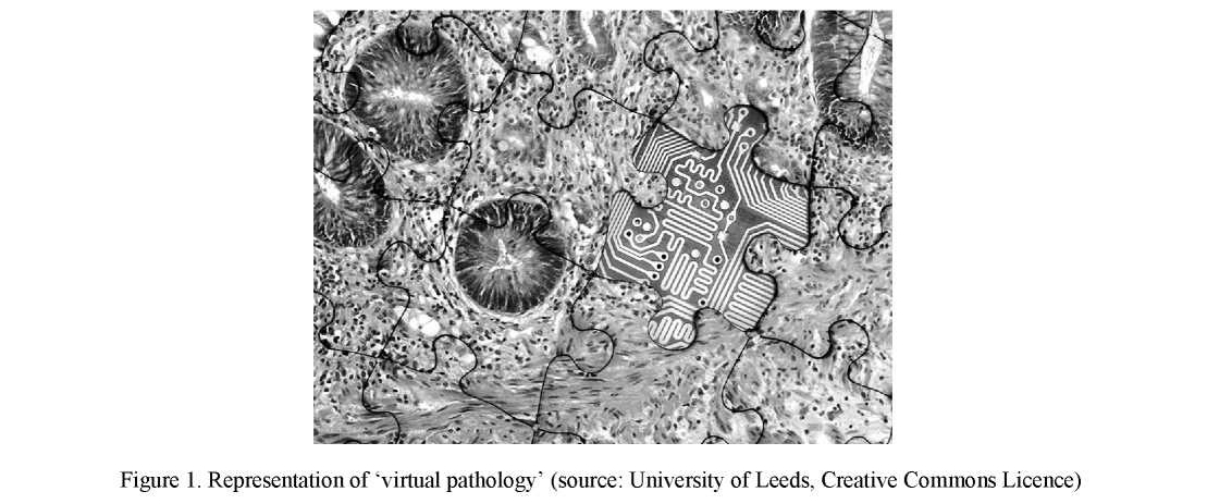
The biggest change in pathology has come about through the advancement of virtual microscopy technology enabling digitization of microscopy slides and presenting new opportunities for digital image analysis. Computerized vision provides an immediate benefit of increased capacity (automation) and precision (reproducibility) [3]. Digital images can be used to make primary diagnoses, offer second opinions (consultation), for telepathology (Fig. 1), quality assurance (e.g. re-review and proficiency testing), archiving and sharing, education and conferencing, image analysis, research and publications, marketing and business purposes, as well as tracking (e.g. audit trail of how an image was viewed).
An example of such a system is a platform provided by Aperio ePathology (there are many others). These systems offer further advantages by allowing workflow is integrated into the institution's overall operational environment. A further example is with the Philips IntelliSite Pathology Solution (PIPS), which is the first whole slide imaging (WSI) system (which refers to scanning of conventional glass slides in order to produce digital slides). Digital slides are used in pathology for education, diagnostic purposes (clinico- pathological meetings, consultations, revisions, slide panels and, increasingly, for upfront clinical diagnostics) and archiving [4]. One challenge with developing WSI platforms is that the file size of the WSI ranges from a few megabytes to several gigabytes, leading to challenges in the area of image storage and management when they will be used routinely in daily clinical practice.
The PIPS uses proprietary hardware and software to scan and digitize conventional surgical pathology glass slides prepared from biopsied tissue at resolutions equivalent to 400 times magnification. These digitized images can then be reviewed and interpreted by pathologists. Technology such as this has considerable educational advantages. For instance, a key skill that histopathologists need to learn is the ability to identify areas of diagnostic relevance from an entire sample. Virtual microscopy will allow this skill to be developed in an Internet-based environment; here the student/trainee can see the morphologic patterns or diagnostic feature in the context of the entire sample. Training on tissue sections would not restricted to being in the same room as the glass slide, and could be provided remotely and virtually from whole slide scans.
Based on the success of digital pathology platforms, the U.S. Food and Drug Administration (FDA) gave approval for such systems to be used with the U.S. in May 2017 [5]. With this, systems are now capable of digitizing slides that would otherwise be stored in physical files. These platforms also provide a streamlined slide storage and retrieval system that may ultimately help make critical health information available to pathologists, other health care professionals and patients faster.
Telepathology
Related to digital imaging is telepathology. Telepathology includes digital microscopy, digital pathology, remote robotic microscopy, teleconferencing, teleconsultation, telemicroscopy, video microscopy, virtual microscopy, web conferencing, and whole slide imaging. Components of a telepathology system include a digital imaging workstation to acquire images, telecommunications network to transmit images, and monitor
or screen to remotely view digital images [6]. Recently telepathology has harnessed the opportunities provided by cloud computing (Fig. 2).
New technologies are being implemented for disease detection; drawing on one example as an illustration a special type of optical fiber, made from a hydrogel, has been developed. This rubber-like device can detect diseases early and send an alert signal. The new device is biocompatible and it can be stretched and pulled in almost any direction. The flexibility comes from the fact the device is compered almost entirely of water, which relates to its hydrogel properties [7].
A hydrogel is a network of polymer chains that are hydrophilic. Such gels are often found as a colloidal gel in which water is the dispersion medium. Hydrogels also possess a degree of flexibility very similar to natural tissue. With this one common use of hydrogels is as scaffolds in tissue engineering. The flexible properties would allow the optical fiber to bend and twist with the natural motions of the body, without breaking. The development brings together the latest research into pliable hydrogels together with new developments in flexible electronics.
The aim is for such a device to be implanted into the body and to either provide an alert about a disease risk, by lighting up, or even to attempt to eliminate certain pathogens by delivering therapeutic pulses of laser light. The success or otherwise would be partly based on the device being located within a specific region of the body. The initial aim is to develop the optical fiber for use in the brain, to provide effective stimulation and therapy. The light-related effects are related to the science of optogenetics. Within this field, light is used to activate cells and there has been considerable success in using light to activate neutrons in the brain. The optical fiber draws its light from micro-sixed LED lights, contained within each strand of the fiber.
Trials are set to begin using the optical fibers for long-term diagnostics, to optically monitor tumors or inflammation in the brain.
To take a different area: histopathology. This is one area that has remained steady, largely reliant upon legacy technology. This is gradually undergoing change and one example of new technology is the Phillip's IntelliSite ultra fast scanner. This is a high-throughput bright-field slide scanner that accommodates current histopathology requirements together with room for expansion. The device also speeds up the time-to-result by allowing preparations to be run overnight. The scanner has a storage capacity of 300 barcoded slides.
Cloud computing
The use of cloud computing as a business tool is well-established. The take-up in the science world has been more varied. As an example of the application, the world's biggest microbial genome project is taking advantage of cloud-based platforms. Cloud computing can be used to analyze microbiological samples. Cloud computing refers to the practice of using a network of remote servers hosted on the Internet to store, manage and process data. It is an alternative to the use of a local server or a personal computer. In a sense the network that forms the cloud can be thought of as an electricity grid. The key advantages are the ability to share data, to enable collaboration, and to offer greater protection of data should a piece of hardware crash.
The University of Warwick has developed a cloud-based microbial bioinformatics resource. The created database is said, by the developers, to be the largest of its type in the world. The project is called the ‘Cloud Infrastructure for Microbial Bioinformatics' (CLIMB) project. The focus is with medical microbiology, where microorganisms that cause disease are analyzed. Social media coverage relating to the CLIMB project are posted via the MRC Climb (@MRCClimb) Twitter feed.
The CLIMB has been set-up through based on the idea of Professor Mark Pallen (University of Warwick). CLIMB represents a user-friendly, one-stop shop for sharing software and data between medical microbiologists in the academic and clinical arenas. The cloud already contains data from dozens of research groups. An important part of the system is virtualization, which enables scientists to work in a simulated computer environment populated by virtual machines. These are placed on top of physical hardware and resemble conventional servers. These provide large data storage capabilities and allow several biological databases to be integrated.
The primary data is taken from molecular sequencing of microbial genomes, which allow medical microbiologists to assess the most appropriate antimicrobials to treat a patient or to track epidemiological trends within the community. The use of such data is termed «bioinformatics.» Bioinformatics is an interdisciplinary field that develops methods and software tools for understanding biological data. Bioinformatics has two aims: the implementation of computer programs to enable efficient access to information; and the creation of new algorithms and statistical tools to understand relationships between items of data within large data sets.
Cellular manipulation
It is important in the context of cell therapies for people to cure specific diseases or regenerate tissues that are no longer functional. This aim has been hampered by technological limitations; however, a new method of cellular manipulation offers a new solution. The research is an example of the use of digital based bio-nanotechnology. The new method can be used for altering the path and direction of cells was developed by Northwestern University, and it has been described in Nature Communications [8]. The aim of the technique is to develop stem cell therapies for spinal cord injuries, stroke, and Parkinson's disease.
The new technology centers on the way that cells behave in the human body. Our cells are continually being signaled with various instructions, triggered by proteins and other molecules that are located in the matrices that surround them. As an example, such signals can be cues for cells to express specific genes in order for the cells to differentiate into other types of cells. Such a development is important for growth or regeneration of tissues. This sophisticated, biological signaling machinery has the pre-programmed capacity to make signals stop and re-start as needed; or to switch off one signal and activate an alternative signal in order to commence a complex processes. If this could be controlled by medics, then the process of addressing a range of diseases could be achieved. So far, the ability to produce such regenerative therapies has proved impossible.
This could be set to change with the development of a synthetic material that can trigger reversibly certain types of signaling. This platform could lead to materials to control stem cells in order to produce regenerative therapies and to control cellular functions.
The new technology should help with research into treatments for such diseases as Alzheimer's disease, Parkinson's disease, problems with arthritic joints, spinal cord injuries, the effects of stroke, and other conditions requiring tissue regeneration.
In trials, the researchers have taken spinal cord neural stem cells (neurospheres) and driven them to differentiate using a signal, helping the scientists to understand developmental and regenerative cues. This cell manipulation technology could help control which cells change and thereby address diseases like Parkinson's, such as converting a patient's own skin cells into stem cells.
Digital colony counting
For microbiologists and pathologists the process of counting bacterial colonies can be tedious and mistakes can happen. Laboratory managers are turning attention to automated, digital devices to streamline processes. Colony counting is the mainstay of many microbiology laboratories. Microbial culture media in the form of semi-solid agar is used to grow up microbial colonies of enumeration. Many microbiological techniques rely on accurate determination of colony forming units (CFUs). For many large laboratories hundreds to thousands of plates require counting each day, after incubation. This is not only repetitious (and arguably a waste of time for employed graduate scientists) it can lead to errors and thus problems of data integrity. In
low count assays minor counting errors will have significant effects. A second type of error is when numbers of CFUs on a plate can lead to false results due to overcrowding of bacteria. Digital technology can help to address these errors (Fig. 3).
Many laboratories have successfully implemented automatic colony counters, and digitalization can address errors introduced during the manual counting process and recording of information and lead to a significant reduction in time taken to analyze colony counting data (Fig. 4). Examples include bioMerieux's EasyCount 2 — EC2 and the ProtoCOL automated counter series. In addition, there is the Whitley aCOLyte (Synbiosis, Cambridge, UK) and the AID BacSpot (AID, Strassberg, Germany).
In terms of functionality, automated colony counters offer:
- Standardized and accurate results. Accuracy is important since colony counting can be affected by numerous parameters related to the physical properties of the colony: size, shape, contrast, and overlapping colonies. To achieve this requires automatic colony separation (for when colonies are positioned close to each other).
- Ability to count colonies within appropriate parameters (such down to 50 microns and measure zones accurately to 0.5 millimeters, within detection limits of 0.1 millimeters).
- Ability to visualize white light and fluorescent colonies.
- The ability to count the entire plate or sectors of the plate.
- Results obtained within one second per plate.
- The display of real-time full-color on-screen images.
- Zoom function for looking at smaller colonies.
- Software to allow for data collection and analysis. Data should ideally be transferrable to a Laboratory Information Management System (LIMS).


The essential elements of automated, digital colony counters include a circular dark field illuminator and a camera with a resolution of 3.3 megapixels or higher (many systems have cameras of higher quality); software with appropriate algorithms; an automated plate holder (with a toolbox to enable communication between the software and the image analyzer). With the software algorithm many work on the basis of A Bayes classifier. This is a simple probabilistic classifier used to study the geometric properties such as ratio between major and minor axis of the group are used to verify the number of colonies contained in the group.
Validation of automated colony counters is important. To ensure the validity of their data, microbiologists need to establish that their automated colony counting method is as accurate as a precise manual count before they implement any new process into their workflow. Weaknesses can occur where there are mixed colonies or, due to inhomogeneity of the agar thickness, discrimination is not possible for all areas of the plate. A further weakness is where confluent growth occurs. The light also needs to be right. These issues can be overcomes as a paper by Brugger and colleagues demonstrates [9]. The researchers found that white light dark field illumination works well but a blue dark field illumination gave the best discrimination of all.
The U.K. experience
As a sign of the importance of the technology the biggest single health employer in the world — England's National Health Service (NHS) — published an overview called «Digital First: Clinical Transformation through Pathology Innovation» [10]. The document describes precisely how healthcare can review and apply new technology to deal with the ever increasing demand for pathology services. The reason for discussing the report briefly here is to emphasise the importance that digital technologies hold for the field of pathology.
The title of the report carries with it the central message, as the text states: «Digital First is focused on harnessing the potential of digital channels to enable patients and healthcare professionals to interact in different ways, reducing face-to-face contact where this is not considered by clinician or patient to be neces- sary.»
While improved turnaround times and greater throughput are central to the NHS driver, the report also highlights the importance of storing digital images, and using these are evidence with any report made by a pathologist. In addition, computerized quantitative analysis can be used for prognostic scores and remote- equipped technology also allows the pathologist to interpret frozen sections some distance away from the laboratory. There are also wins for the patient, according to the report in terms of data access. Here people will feel more in control of their health through better access to test results [11].
Reasons for the NHS promoting the digital message include the advantages for improving communications, procedures, workload and quality. There are other advantages too, which Digital Journal has explored in a companion article titled «Pathology services are embracing digital technology.»
Conclusion
This chapter has considered some of the developments that have taken place with and which are shaping digital pathology. Of the different technologies surveyed, digital imaging is the one that will be adopted at the fastest rate. Today software programs enable pathologists to create and read the digital «slides.» Unlike traditional histological samples, microscopic users are able to create digital images of live and dead tissues; the wide range of slide possibilities also includes positive and negative gram stains, blood smears, animal, and plant cells — almost any tissue sample traditionally viewed under a microscope. The advantages offered by cloud computing also allow researchers to create accessible databases and to share images rapidly.
In outlining current trends, the chapter has described many of the advantages that arise from digitalization. The process remains a developing arena, however and there remain barriers to adoption. Barriers to adoption include limiting technology, image quality, problems with scanning all materials (e.g., cytology slides), cost, digital slide storage, inability to handle high-throughput routine work, regulatory barriers, ergonomics, and pathologists' reluctance. In time, many of these will be overcome as technology and education move on.
With barriers slipping, what might the future of digital pathology look like? Some possibilities are:
- Providing primary diagnosis of disease.
- Facilitating personalized medicine where therapies are tailored to the individual.
- Extracting and analyzing data to understand the links between tests and treatments, and to maximize outcomes.
- Helping pathologists access prior data and data from a spectrum of different data sites quickly and easily.
- Providing pathologists with a complete view of patients' health and care.
- Providing an optimized delivery structure by removing geographical boundaries and limitations.
- Empowering people to manage their own health through access to electronic health records.
- Enabling pathologists to interact more easily with patients directly.
How quickly these come to pass remains to be seen; the key message is, however, that digital technology is transforming healthcare and the future delivery of pathology services in new and remarkable ways.
References
- Mistry N. Endoscope: an innovation in mobile endoscopic technology transforming the delivery of patient care in otolaryngology / N. Mistry, C. Coulson, A. George // Expert Rev. Med. Devices. — 2017. — No. 14(11). — P. 913–918.
- Farahani N. Whole slide imaging in pathology: advantages, limitations, and emerging perspectives / N. Farahani, A.V. Parwani, L. Pantanowitz // Pathology and Laboratory Medicine International. — 2014. — № 7. — P. 23–33.
- Pinco J. Impact of digital image manipulation in cytology / J. Pinco, R.A. Goulart, C.N. Otis, J. Garb, L. Pantanowitz // Arch Pathol. Lab. Med. — 2009. — No. 133(1). — P. 57–61.
- Al-Janabi S. Digital pathology: current status and future perspectives / S. Al-Janabi, A. Huisman, P.J. Van Diest // Histopathology. — 2012. — No. 61(1). — P. 1–9.
- FDA (2017) allows marketing of first whole slide imaging system for digital pathology, US Food and Drug Administration. www.fda.gov. Retrieved from https://www.fda.gov/NewsEvents/Newsroom/PressAnnouncements/ucm552742.htm.
- Weinstein R.S. Overview of telepathology, virtual microscopy, and whole slide imaging: prospects for the future / R.S. Weinstein, A.R. Graham, L.C. Richter et al. // Human Pathology. — 2009. — No. l40(8). — P. 1057–1069.
- Guo J. Highly Stretchable, Strain Sensing Hydrogel Optical Fibers / J. Guo, X. Liu, N. Jiang et al. // Advanced Materials. — 2016. — No. 28(46). — P. 10244–10249.
- Freeman R. Instructing cells with programmable peptide DNA hybrids / R. Freeman, N.Stephanopoulos, Z. Alvarez et al. // Nature Communications. — 2017. — No. 10(8).
- Brugger S.D. Automated Counting of Bacterial Colony Forming Units on Agar Plates, PLoS One, 2012 / S.D. Brugger, C. Baumberger, M. Jost // Retrieved from at: https://doi.org/10.1371/journal.pone.0033695.
- National health Service «Digital First: Clinical Transformation through Pathology Innovation», National Pathology Programme, NHS, London // Retrieved from https://www.england.nhs.uk/wp-content/uploads/2014/02/pathol-dig-first.pdf (accessed 20th September 2017)
- Armstrong-Smith I. Facing the Digital Future of Pathology / I. Armstrong-Smith // The Pathologist. — 2017. — Iss. 114. — P. 301–302.