In the experiment on CBA mice the effect of lithium carbonate on the liver structure when used as an antitumor agent was studied. Tumor growth was modeled by introducing hepatocarcinoma-29 cells into the muscle tissue of the right thigh of experimental animals. Lithium carbonate was administered on the periphery of tumor growth at a dose of 0.037 mg per animal. The structure of the liver was studied by light, electron microscopy and morphometry after 3, 7, 13, and 30 days of the experiment. In the dynamics of remote tumor growth, structural changes in the liver were discovered, which indicates the organ dysfunction. It has been shown that by the 30th day of tumor development there was a decrease in the volume and number density of hepatocytes, a decrease in the number density of binuclear cells and an increase in the volume density of sinusoids. In hepatocytes there was a decrease in the concentration of all intracellular organelles, which is a reflection of a decrease in the energy and synthetic function of cells. The use of lithium carbonate as an antitumor agent led to the aggravation of structural changes in the liver, which was apparently due to an increase in the toxic load on the organ due to an increase in the decay products and the dying of tumor cells under the influence of lithium.
Introduction
Hepatocarcinoma is one of the most aggressive human tumors and, despite advances in early diagnosis and treatment, leads to high mortality rate among patients due to metastasis. [1]. It is known that lithium compounds (lithium chloride and lithium carbonate) can influence the signaling pathways and cell cycle regulation [2], and possess immunomodulating properties [3]. There is research showing the effectiveness of the use of lithium compounds to suppress tumor growth [4-6].
The liver is a parenchymal organ and at the same time it is the largest gland of internal secretion of a complex tubular structure. Its main role is the formation and secretion of bile, which is involved in the conversion of fatty acids into soluble compounds that can be absorbed in the digestive tract. The following processes occur in liver: glycogen synthesis and deposition, its reverse transformation into sugar and discharge into the blood as the body needs. [7]. The study of the fine structure and nature of the vital activities of these cells in the normal condition, as well as the changes that occur in them under various influences, is of great practical interest [8]. Complex and diverse functions of the liver is provided by the work of the cellular elements of its parenchyma — hepatocytes [9].
The use of new approaches to the study of the influence of malignant growth and anticancer drugs on the structural and functional state of the liver is topical.
Research objective
Identifying the effect of lithium carbonate on the structural organization of the liver when it is used for the correction of peripheral tumor growth.
Material and methods of research
An experimental study was carried out on CBA mice weighing 18–20 g at the age of 3 months. The animals were kept on a standard diet with free access to water and food. Work with animals was carried out in accordance with the «Rules of work with the use of experimental animals».
In the experiment there were 3 groups of animals. Group 1 consisted of intact animals, group 2 consisted of animals with the development of a tumor process (n = 20), group 3 consisted of animals that, after induction of a tumor process, received injections of lithium carbonate into the right thigh (n = 20). Hepatocarcinoma-29 (HC-29) cells were used to induce tumor growth. HC-29 was obtained and verified by the staff of the Institute of Cytology and Genetics of the Siberian Branch of the Russian Academy of Sciences [10] and was kindly provided for our research. HC-29 cells were transferred to CBA mice's abdominal cavity, after 10 days, ascitic fluid was taken and suspended in 10-fold volume of physiological saline and injected in a volume of 0.1 ml into the muscle of the right thigh of intact animals. Animals in group 3 received five intramuscular injections in the right thigh in a volume of 0.1 ml of lithium carbonate suspension prepared with sterile 0.85 % aqueous solution of sodium chloride in a dose of 0.037 mg per animal. Intramuscular administration of lithium carbonate was done to simulate the process of drug delivery to the site of implantation of tumor cells. The material was taken for research after 3, 7, 13, and 30 days of the experiment. Animals were drawn out of the experiment under ether anesthesia by the method of cranio-cervical dislocation. For electronic and microscopic examination, liver samples were fixed in 4 % paraformaldehyde solution prepared on Hanks medium, fixed for 1 hour in 1 % OsO4 solution (osmium tetroxide) (Sigma, USA)on phosphate buffer (pH = 7.4), dehydrated in ethanol of increasing concentration and put in epon (Serva, Germany). Semi-thin sections with a thickness of 1 μm were obtained on a Leica EM UC7 ultramicrotome (Germany / Switzerland), stained with toluidine blue, studied under a LEICA DME light microscope (Germany), photographed using the Avigion computer program. Photomicrographs were measured morphometrically using the ImageJ computer program (USA). Volume density of the parenchyma and stroma of the liver, the number density of hepatocytes and their nuclei using a closed test system of 120 points were estimated. Ultrathin sections of 70–100 nm thick were contrasted with a saturated aqueous solution of uranyl acetate and lead citrate and examined in a JEM 1010 electron microscope (Japan). Hepatocytes were morphometrically measured using the ImageJ computer program. The volume density of mitochondria, cisterns of the rough-surface endoplasmic reticulum, lipid vacuole, glycogen, primary, secondary lysosomes and autophagolysosomes were evaluated. Statistical data processing was performed using the program Statistica 6.0. The mean values and the standard deviation were calculated, the significance of differences was calculated using the Mann-Whitney U-test and was taken at p <0.05.
Results and discussion
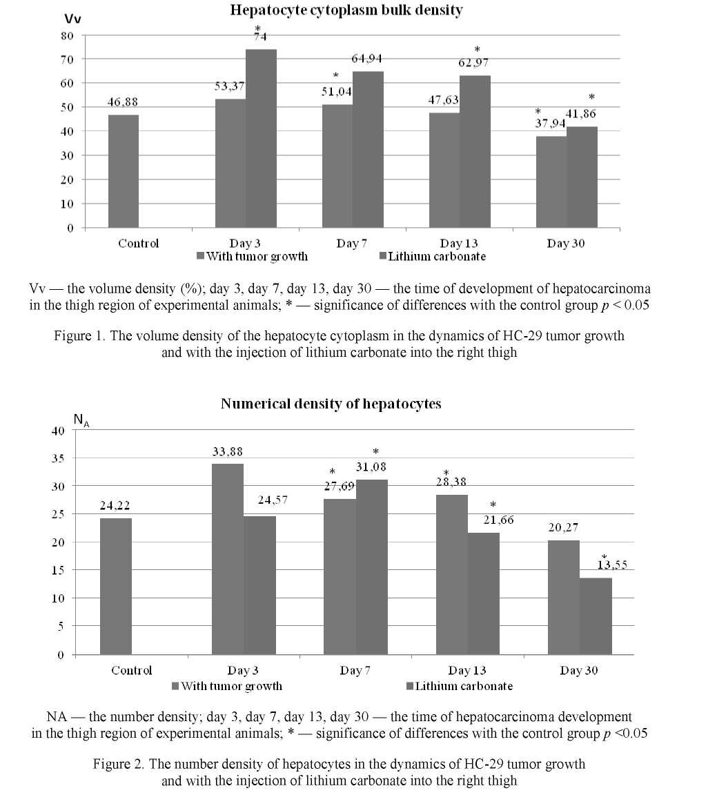
On day 30 of the experiment under conditions of remote tumor growth, the volume density of the cytoplasm of hepatocytes of animals with tumor growth was 11 % less (p <0.05) than in control animals (Fig. 1). At the same time, the value of the hepatocyte number density decreased by 30 % (p <0.05) (Fig. 1b) during the experiment.
In animals which received lithium carbonate injections in the right thigh, an increase in the volume density of the cytoplasm of hepatocytes was observed from day 3 to day 13 of the study. On day 3, the value of this indicator was increased by 13 %, on day 7 — by 9 % and on day 13 — by 1.5 % compared with the corresponding value in the control group (Fig. 1). By day 30 of the study, the volume density of the hepatocyte cytoplasm did not differ from the level in the control group (Fig. 1).
On day 3, day 7 and day 13 of remote tumor growth, there was an increase in the number density of hepatocytes by 40 %, 14 %, and 18 % (p < 0.05), compared with the control group (Fig. 2). The increase in the hepatocyte number density in animals treated with lithium carbonate was observed on day 7, and a decrease was observed by days 13 and 30 by 10 % and 40 % respectively (p <0.05) (Fig. 2).
In animals with tumor growth, there was a decrease in the volume density of sinusoids on all days of the experiment, on day 3 by 18 %, on day 7 by 10 % (p <0.05), on day 13 by 3 % (p < 0.05), and on day 30 the value of this parameter decreased by 3 times, compared with the animals of the control group (Fig. 1). In animals treated with lithium carbonate, a decrease in the sizes of sinusoids was also observed on day 7 by 35 % (p <0.05), on day 13 by 2 % (p <0.05), and on day 30 the sizes decreased by 6 times (Fig. 3).
A morphometric analysis of the number density of hepatocyte nuclei in the dynamics of tumor growth revealed their significant change on day 3 of the experiment, the value of this indicator increased by 56 % (p < 0.05), compared with animals of the control group (Fig. 4). On day 30 of the experiment the indicator decreased by 19 % compared with that of day 3 (p < 0.05). In animals that received injections of lithium carbonate in the right thigh, the value of the number density of hepatocyte nuclei of animals with tumor growth increased by 56 % (p < 0.05) on day 3, and on day 30 there was a decrease in the number density of hepatocyte nuclei by 2 times in comparison with that of day 7 (Fig. 4).
During the study of binuclear hepatocytes, there was a decrease by 10 times and 3.5 times (p <0.05) on day 30 of the experiment with tumor growth and the introduction of lithium carbonate, respectively (Fig. 5).


The lymphatic drainage of hepatocytes is associated with non-vascular microcirculation and consists of the Disse's space and periacinal Malla's spaces [11]. During the morphometric analysis of Disse's spaces in the dynamics of tumor growth a significant change in their sizes was found. After 7 days of the experiment, there was an increase in the sizes of Disse's spaces by 28 % (p < 0.0001), and after 13 days the value of this parameter began to decrease from the values of day 7 by 20 % (p < 0.0001), and by day 30 the value of this parameter decreased by 2 times (p < 0.0001). By day 30 of the experiment, the value of this parameter returned to its original value (Fig. 6). In animals treated with lithium carbonate injections in the right thigh, the sizes of Disse's spaces decreased by 18 % (p < 0.0001), on day 3, compared with the values in the control group, and from day 3 to day 13 of the study, there was a sharp decrease in these parameters (Fig. 6). On day 13 the indicator of the sizes of Disse's spaces decreased by 9 times, and on day 30 its value increased by 74 % (p <0.0001), compared to day 13 of the experiment (Fig. 6).
A change in the ultrastructural organization of hepatocytes was found. The main organelles that provide the cell with energy are mitochondria. In the liver of animals with tumor growth, a decrease in the volume density of mitochondria was observed (Fig. 7). Mitochondria are cellular organelles that play an important role in bioenergetic processes. Their function leads to providing the cell with chemical energy.


On day 3, the value of this indicator decreased by 45 % (p < 0.05), and on day 30 of tumor growth it was 68 % (p < 0.05) of the corresponding value in the control group (Tables 1, 2). When exposed to lithium carbonate, a decrease in the volume density of mitochondria was observed for all study periods. On day 3, the value of this indicator was reduced by 45 %, on day 7 — by 9 % (p < 0.05), and on day 13 — by 7 % (p < 0.05) compared with the corresponding value in the control group (Tables 1, 2).
There was an increase of volume density of lysosomal structures in cells, in particular autophagolysosomes. The value of this indicator increased by 2 times by day 30 of the experiment (Tables 1, 2). At the same time, all stages of intracellular autophagic degradation were noted: the presence of hepatocytes autophagosomes with organelles in the cytoplasm (Table 1). In autophagosomes, fragments of cytoplasm, glycogen rosettes, mitochondria, fragments of the endoplasmic reticulum with ribosomes were observed.
During the first 7 days of tumor development in the muscle tissue of the thigh in the cytoplasm of hepatocytes, there was a tendency of decrease in the volume density of lipid vacuoles. On days 13 and 30 of the experiment, the value of this indicator decreased by 4.5 and 3 times (p < 0.05) respectively (Table 1). The volume density of glycogen decreased by 3 times (p < 0.05) on day 3 of tumor development, subsequently, the value of this indicator did not significantly differ from the corresponding value in the control group (Tables 1, 2). On day 13, experimental animals that received injections of lithium carbonate in the right thigh showed a tendency of increase of lysosomal structures by 2.5 times (p < 0.05) relative to day 13, and on day 30 it decreased by 45 % (Tables 1, 2).
Table 1 The results of a morphometric analysis of the ultrastructural organization of the liver under conditions of remote tumor growth with the introduction of lithium carbonate, days 3 and 7 of research (M ± SD)
Note. * — significance of differences with control group p < 0.05; 1 — significance of differences with day 3 LC p < 0.05; # — significance of differences with day 7 LC p < 0.05; 0 — significance of differences with day 13 LC p < 0.05.
|
Parameters |
Control |
Day 3 TG |
Day 3 LC |
Day 7 TG |
Day 7 LC |
|
Hepatocyte cytoplasms, Vv |
70.01 ± 1.34~~ |
68.24 ± 2.4^3~ |
13.96 ± 2.2^8~ |
66.18 ± 0.64*~ |
8.09 ± 0.94~~ |
|
Hepatocyte, NA |
34.15 ± 0.72 |
33.92 ± 1.22 |
27.59 ± 0.72 |
||
|
Mitochondria, Vv |
37.52 ± 1.17 |
29.22 ± 1.01 |
20.75 ± 3.42**~ |
34.22 ± 1.24 |
14.94 ± 2.27*1~ |
|
Granular SER, Vv |
33.63 ± 2.45 |
38.74 ± 1.30 |
21.85 ± 3.58 |
31.2 ± 1.32 |
11.08 ± 1.18 |
|
Agranular SER, Vv |
6.66 ± 0.41 |
5.46 ± 0.11 |
3.76 ± 0.61 |
7.35 ± 0.56 |
7.12 ± 0.87 |
|
Golgi apparatus, Vv |
2.60 ± 0.21 |
3.56 ± 0.42 |
3.17 ± 0.52 |
2.55 ± 0.23 |
7.25 ± 0.78 |
|
Attached ribosomes, N A |
85.75 ± 3.63 |
73.23 ± 2.46 |
13.6 ± 2.25 |
78.16 ± 3.56 |
26.47 ± 1.25 ι |
|
Free polysomal ribosomes, NA |
40.12 ± 0.99 |
22.24 ± 0.64 |
13.76 ± 2.25 |
13.22 ± 1.51* |
28.55 ± 3.03^*1~ |
|
Autophagosomes, Vv |
0.02 ± 0.01 |
0.48 ± 0.39 |
0 ± 0 |
1.38 ± 0.19 |
6.46 ± 1.02 |
|
Primary lysosomes, Vv |
2.00 ± 0.11 |
2.67 ± 0.47 |
2.41 ± 0.39* |
1.72 ± 0.05* |
SK SK SK 6.18 ± 0.74 * |
|
Secondary lysosomes, Vv |
1.06 ± 0.11 |
3.88 ± 0.64 |
2.19 ± 0.63 |
1.61 ± 0.10 |
3.26 ± 0.41 ι |
|
Lipid vacuole, Vv |
8.00 ± 0.71 |
6.21 ± 0.65 |
21.28 ± 3.48 |
6.11 ± 0.31 |
6.84 ± 1.05**τ~ |
|
Glycogen, Vv |
14.21 ± 0.59 |
4.71 ± 1.02 |
8.71 ± 1.43 |
16.72 ± 1.19 |
13.84 ± 1.49*πr |

Table 2
The results of a morphometric analysis of the ultrastructural organization of the liver under conditions of remote tumor growth with the introduction of lithium carbonate, day 13 and day 30 of the study (M ± SD)
|
Parameters |
Control |
Day 13 TG |
Day 13 LC |
Day 30 TG |
Day 30 LC |
|
Hepatocyte cytoplasms, Vv |
70.01 ± 1.34~ |
65.35 ± 0.50* |
11.07 ± 1.8~ |
55.45 ± 1.58*~ |
12.37 ± 1.92 |
|
Hepatocyte, NA |
34.15 ± 0.72 |
28.38 ± 0.44 |
20.27 ± 0.69 |
||
|
Mitochondria, Vv |
37.52 ± 1.17 |
34.78 ± 0.58 |
18.58 ± 3.04**~ |
25.17 ± 0.87 |
20.30 ± 3.14 |
|
Granular SER, Vv |
33.63 ± 2.45 |
30.15 ± 1.80 |
16.19 ± 2.65 |
20.18 ± 0.77 |
16.98 ± 2.71 ʌ |
|
Agranular SER, Vv |
6.66 ± 0.41 |
7.09 ± 0.32 |
17.28 ± 2.83#~ |
2.90 ± 0.19 |
9.72 ± 1.62000 |
|
Golgi apparatus, Vv |
2.60 ± 0.21 |
2.09 ± 0.16 |
3.62 ± 0.59 |
3.19 ± 0.27 |
2.49 ± 0.39 |
|
Attached ribosomes, N A |
85.75 ± 3.63 |
77.40 ± 3.62 |
12.30 ± 2.0Īɪ¯ |
29.36 ± 1.34, |
22.17 ± 3.58 |
|
Free polysomal ribosomes, NA |
40.12 ± 0.99 |
10.46 ± 0.83 |
20.81 ± 3.47 |
11.54 ± 0.36 |
17.55 ± 2.95*"00 |
|
Autophagosomes, Vv |
0.02 ± 0.01 |
0.84 ± 0.10 |
1.46 ± 0.24 , |
2.68 ± 0.15 |
13.30 ± 2.1200 |
|
Primary lysosomes, Vv |
2.00 ± 0.11 |
3.15 ± 0.20 |
3.33 ± 0.54 |
2.73 ± 0.15 |
2.80 ± 0.45 |
|
Secondary lysosomes, Vv |
1.06 ± 0.11 |
1.78 ± 0.11 |
5.0 ± 0.82* |
2.37 ± 0.14* |
2.75 ± 0.44** |
|
Lipid vacuole, Vv |
8.00 ± 0.71 |
1.78 ± 0.1Γ |
12.93 ± 2.11" |
2.37 ± 0.40* |
6.19 ± 1.04**0 |
|
Glycogen, Vv |
14.21 ± 0.59 |
13.5 ± 0.51 |
9.35 ± 1053# |
17.75 ± 0.91*~ |
0 ± 0*000 |
Note. * — significance of differences with control group p < 0.05; 1 — significance of differences with day 3 LC p < 0.05; # — significance of differences with day 7 LC p < 0.05; 0 — significance of differences with day 13 LC p < 0.05.
Structural changes in the liver were determined by the development of tumor in the muscle tissue of the thigh. During the implantation of hepatocarcinoma-29 cells into the thigh region of experimental animals, tumor cells quickly filled the intermuscular spaces. By day 13 of the experiment, they replaced the muscle fibers, forming a tumor node, and by day 30 after the implantation, the tumor cells formed a kind of liver beams, surrounded by «sinusoids» [12]. At the same time, significant changes in free-radical lipid oxidation were observed in the dynamics of tumor growth. There was an increase in the level of secondary products of lipid peroxidation (LPO) in muscle tissue: on day 7 — 2.1 times, on day 13 — 1.4 times relative to control values, which was associated with invasion of tumor cells and damage to membrane structures [13, 14] and could not have a toxic effect on the liver. With the introduction of lithium carbonate, the destruction of tumor cells and the concentration of lipid peroxidation products significantly increased [11, 13, 14].
Currently, the main medical indications for using lithium are aimed at the treatment of acute bipolar disorder (BD) and for adjuvant treatment of major depression, given its well-proven mood stabilization properties [15]. The effectiveness of using lithium for the treatment of prostate cancer was shown [16].
Lithium salts are considered as a potential therapeutic target for numerous human malignant tumors. Many different effects of lithium have been found by researchers studying various types of cells, organs and organisms. However, the difficulty lies in determining which of the mechanisms of action of lithium are important for its therapeutic effect [17].
Our findings suggest that the biological effects of lithium on the structure of the liver are primarily due to its effect on hepatocarcinoma cells. Stimulating the destraction of tumor cells, lithium carbonate contributes to the increase of toxic products of decomposition of tumor tissue, thereby increasing the load on the organs of detoxification, in particular, the liver.
Conclusion
Using light and electron microscopy, it was revealed that under conditions of remote tumor growth, structural changes develop in the liver, indicating a violation of the function of the organ. The volume and number density of hepatocytes, also the number density of binuclear hepatocytes decrease, reflecting impaired hepatocyte renewal processes under the toxic effects of tumor metabolism products. In hepatocytes, the concentration of intracellular organelles decreases, which is a reflection of the decrease in the energy and synthetic function of cells. The use of lithium carbonate as an antitumor agent led to the aggravation of structural changes in the liver, which was apparently caused by the increase in the toxic load on the organ due to an increase in the decay products and the destruction of tumor cells under the influence of lithium.
This work was supported by the grant of JSC «Center for International Programs» Contract No. 4141, dated December 26, 2016 and budgetary funding RICEL — branch of ICaG SB RAS № 0324-2019-045-С- 02.
References
- Capece, D., Fischiett, M., & Versella, D., et al. (2013). Biomed Research International, 2013, 187204, 15. http://doi.org/ 10.1155/2013/187204
- Berasain, C., Castillo, J., & Perugorria, M.J. et al. (2009). Ann. NY Acad Sci., 1155, 206-2011
- Wang, J.S., Wang, C.L., & Wen, J.F., et al. (2008). Lithium inhibits proliferation of human esophageal cancer cell line Eca- 109 by inducing a G2/M cell cycle arrest. World J. Gastroenterol., 14, 25, 3982-3989.
- Raghavendra, P.B., Lee, E., & Parameswaran, N. (2013). J. Neuroimmune Pharmacol., Nov. 27.
- Sun, A., Shanmugam, I., & Song, J., et al. (2007). Lithium suppresses cell proliferation by interrupting E2F-DNA interaction and subsequently reducing S-phase gene expression in prostate cancer. Prostate, 67, 976-988.
- Zhu, Q., Yang, J., & Han, S., et al. (2011). Suppression of glycogen synthase kinase 3 activity reduces tumor growth of prostate cancer in vivo. Prostate, 71, 8, 835-845.
- Vrakin, V.F., & Sidorova, M.V. (1984). Anatomiia i ^stolohlia Clomashnii ptitsy [Anatomy and Histology of Domestic Fowl]. Moscow: Kolos [in Russian].
- Goralskiy, L.P., Khomich, V.T., Kot, T.F., & Guralska, S.V. (2011). Anatoimia svπskikh ptaklnv [Anatomy of Domestic Fowl]. Zhitomir: Pohssia [in Russian].
- Usha, B.V. (1979). Strukturnaia orhanizatsiia pecheni v usloviiakh termicheskoho ozhoha kozhi i korrektsii [Structural organization of the liver under conditions of thermal skin burn and correction]. Veterinarnaya hepatolohiia — Veterinary Hepatology. Moscow: Kolos [in Russian].
- Kaledin, V.I., Zhukova, N.A., & Nikolin, V.P., et al. (2009). Hepatokartsinoma-29 — metastaziruiushchaia perevivaemaia opukhol myshei, vyzyvayushchaia kakheksiiu [Hepatocarcinoma-29 — metastatic transplantable tumor of mice causing cachexia]. Biulleten eksperimentalnoi biolohii — Bulletin of Experimental Biology, 148, 12, 664-669 [in Russian].
- Borodin, Yu.I., Michurina, S.V., Ishchenko, I.Yu., & Belkin, A.D. (2012). Limfaticheskii rehion i hematolimfaticheskii barer pecheni v norme [Lymphatic region and liver hematolymphatic barrier in normal condition]. Limfolohiia — Limfology. (Eds. V.I. Konenkov, Yu.I. Borodin, M.S. Lyubarskii). Novosibirsk: Manuskript [in Russian].
- Bgatova, N.P., Borodin, Yu.I., Makarova, V.V., Pozhidayeva, A.A., Rachkovskaya, L.N., & Konenkov, V.I. (2014). Vliianie nanorazmernykh chastits karbonata litiia na intaktnuiu myshechnuiu tkan i opukholevyi rost [The effect of nanosized lithium carbonate particles on intact muscle tissue and tumor growth]. Biulleten eksperimentalnoi biolohii — Bulletin of Experimental Biology, 157, 1, 102-108 [in Russian].
- Konenkov, V.I., Borodin, Yu.I., Makarova, O.P., Bgatova, N.P., & Rachkovskaya, L.N. (2015). Effekty nanorazmernykh chastits karbonata litiia na oksidantno-antioksidantnyi status opukholevoi tkani pri razvitii hepatokartsinomy-29 [Effects of nanosized particles of lithium carbonate on the oxidative-antioxidant status of tumor tissue during the development of hepatocarcinoma- 29]. Patolohicheskaia fiziolohiia i eksperimentalnaia terapiia — Pathological physiology and experimental therapy, 59, 2, 57-64 [in Russian].
- Bgatova, N.P., Makarova, O.P., Pozhidayeva, A.A., Borodin, Yu.I., Rachkovskaya, L.N., & Konenkov, V.I. (2014). Effects of Lithium Nano-Scaled Particles on Local and Systemic Structural and Functional Organism Transformations Under Tumour Growth. Achievements in the Life Sciences, 8, 2, 101-111.
- Nivoli, A.M., Kolom, F., Murru, A., Pacchiarotti, I., Kastro-Loli, P., Gonsales-Pinto, A. et al. (2011). Novye rukovodiashchie printsipy lecheniia ostroi bipoliarnoi depressii: sistematicheskii obzor [New guidelines for the treatment of acute bipolar depression: a systematic review]. Affektivnye rasstroistva — Affective disorders, 129 (1–3), 14-26 [PubMed] [Google Scholar] [in Russian].
- Hossein, G.Z., Avareh, V.A., & Fard, P.S. (2012). Combined Treatment of Androgen-Independent Prostate Cancer Cell Line DU145 with Chemotherapeutic Agents and Lithium Chloride: Effect on Growth Arrest and/or Apoptosis. Avicenna J. Med. Biotechnol., Apr. 4(2), 75-87.
- Alda M. (2015). Lithium in the treatment of bipolar disorder: pharmacology and pharmacogenetics. Mol Psychiatry, Jun. 20(6), 661-670. doi: 10.1038/mp.2015.4. Epub 2015 Feb 17.